
Radiography (X-Ray)
Radiography is the imaging of body structures, or parts of the body, using X-rays. X-rays are a form of radiation (X-radiation) similar to visible light, radio waves and microwaves. X-radiation is special because it has a very high energy level that allows the X-ray beam to penetrate through the body and create an image or picture.
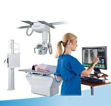
The HF advantage high frequency X-ray unit from GE healthcare is an evolution to the next level in X-ray technology. High Frequency X-ray technology produces crystal clear images at the lowest radiation dose possible, surpassing conventional X-ray performance. It typically reduces exposure time by 30%, lowers radiation dose by 25%. This high frequency technology is complemented by the addition of computed radiography, enabling exceptional Digital X-ray images.
How are X-rays performed?
X-rays can be performed on an outpatient basis, or as part of inpatient care.
Although each facility may have specific protocols in place, generally, an X-ray procedure follows this process:
- The patient will be asked to remove any clothing or jewellery which might interfere with the exposure of the body area to be examined. The patient will be given a gown to wear if clothing must be removed.
- The patient is positioned on an X-ray table that carefully positions the part of the body that is to be X-rayed–between the X-ray machine and a cassette containing the X-ray film or specialized image plate. Some examinations may be performed with the patient in a sitting or standing position.
- Body parts not being imaged may be covered with a lead apron (shield) to avoid exposure to the X-rays.
- The X-ray beam will be aimed at the area to be imaged.
- The patient must be very still or the image will be blurred.
- The technologist will step behind a protective window and the image is taken.
- Depending on the body part under study, various X-rays may be taken at different angles, such as the front and side view during a chest X-ray.
Why it’s done
- X-ray of knee arthritis
- Chest X-ray
- Bones and teeth
- Fractures and infections. In most cases, fractures and infections in bones and teeth show up clearly on X-rays
- Arthritis. X-rays of your joints can reveal signs of arthritis. X-rays taken over the years can help your doctor determine if your arthritis is worsening.
- Dental decay. Dentists use X-rays to take pictures of the teeth and jaw and check for cavities.
- Osteoporosis. Special types of X-ray tests can measure bone density.
- Bone cancer. X-rays can reveal bone tumors.
Chest
- Lung infections or conditions. Evidence of pneumonia, tuberculosis or lung cancer can show up on chest X-rays.
- Breast cancer.Mammography is a special type of X-ray test used to examine breast tissue.
- Enlarged heart. This sign of heart failure shows up clearly on X-rays.
- Blocked blood vessels. Changes in blood flow to the lungs and heart can be seen on chest X-rays.
Abdomen
- Digestive tract problems. Barium, a contrast medium delivered in a drink or an enema, can help reveal problems in your digestive system.
- Swallowed items. If your child has swallowed something like a key or a coin, an X-ray can show the location of that object.
Radiation exposure
Some people worry that X-rays aren’t safe because radiation exposure can cause cell mutations that may lead to cancer. The amount of radiation you’re exposed to during an X-ray depends on the tissue or organ being examined. Sensitivity to the radiation depends on your age, with children being more sensitive than adults.
Generally, however, radiation exposure from an X-ray is low, and the benefits from these tests far outweigh the risks.
However, if you’re pregnant or suspect that you may be pregnant, tell your doctor before having an X-ray. Though the risk of most diagnostic X-rays to an unborn baby is small, your doctor may consider another imaging test, such as ultrasound.
After the X-ray
After an X-ray, you generally can resume normal activities. Routine X-rays usually have no side effects. However, if you receive contrast medium before the test, drink plenty of fluids to help rid your body of it.